0:00
Introduction
In part I of this series, we saw how the meniscus, or semilunar cartilage, went from being viewed as a functionless remnant to a critical structure in the health and stability of the knee. At the time, surgery was done open, through an incision in the front of the knee large enough to dissect the meniscus from its capsular attachments. The decision for surgery was also based almost exclusively on history and physical examination.
The physician would listen to the story of the injury, examine the knee, such as with a McMurray type maneuver, and decide on surgery. Sources from this time discuss their hit rate for correct diagnosis at the time of surgery, some stating they made the correct diagnosis one hundred percent of the time. Granted, the surgeon removing the meniscus was also the one recording the diagnosis, and that diagnosis was confirmed only after the meniscus had already been cut out.
This chapter traces the remarkable period when meniscus care moved from indirect diagnosis and open surgery toward clearer visualization, smaller incisions, and a deeper respect for preservation. From arthrography and Watanabe’s arthroscope to the science of healing zones and MRI, these decades reshaped how surgeons understood, repaired, and protected the meniscus.
The Advent of Advanced Visualization in the 1950s – 1980s
1950s, 1960s
Advances in Imaging: Arthrography
Freiberger RH, Killoran PJ, Cardona G. Arthrography of the knee by double-contrast method. Am J Roentgenol. 1966;97(3):736-747.
Before arthroscopy, diagnosing meniscus tears was primarily done with a clinical exam showing a locking knee, effusion, and negative radiographs. By the early twentieth century, contrast arthrography had become the first effective imaging technique for meniscal injuries. For the first time, surgeons had something more than the patient’s story, the physical exam, and an educated guess.
In the 1930s, clinicians began injecting air or contrast dye into the knee and taking X-rays to visualize the menisci. Over the next two decades, techniques improved. By the 1950s and 1960s, double contrast knee arthrography, using both air and iodine contrast, was the gold standard radiologic test for a suspected meniscal tear. A well performed arthrogram would show filling defects where a torn meniscus disrupted the contrast outline.
Primitive by modern standards, arthrography was still hugely important. It allowed many tears to be confirmed without open surgery. That meant more targeted interventions and a better understanding of tear patterns. Arthrograms remained in use through the 1970s as the mainstay of meniscus imaging, until the advent of MRI in the 1980s rendered them largely obsolete.
Interestingly, reports around this time cite deaths due to large volumes of air being injected into the body, causing air embolisms. The practice of using air as a contrast medium, and these reports, are likely the origin of the misunderstood myth that a small injection of air into the body is dangerous. It is an interesting side story in the history of the meniscus.
“Air and the other more quickly absorbed gases are still being used. Their main disadvantage is that with the large quantities which are used, there is uncomfortable distention of the knee as well as the possible hazard of air embolism. One death and one near death from this cause have been reported. The difference in density between the relatively radiolucent cartilage and the surrounding air is not very great and interpretation of roentgenograms can be difficult. Despite previous aspiration, joint effusion may reduce diagnostic accuracy, apparently because the gas does not always displace the residual fluid within meniscal tears.”
1960s
Masaki Watanabe and the Advent of Arthroscopy
World’s First Meniscectomy – Watanabe 1962. ISAKOS Archive Video (special presentation, 1995). International Society of Arthroscopy, Knee Surgery and Orthopaedic Sports Medicine (ISAKOS). Available via Global Link/ISAKOS membership portal or YouTube.
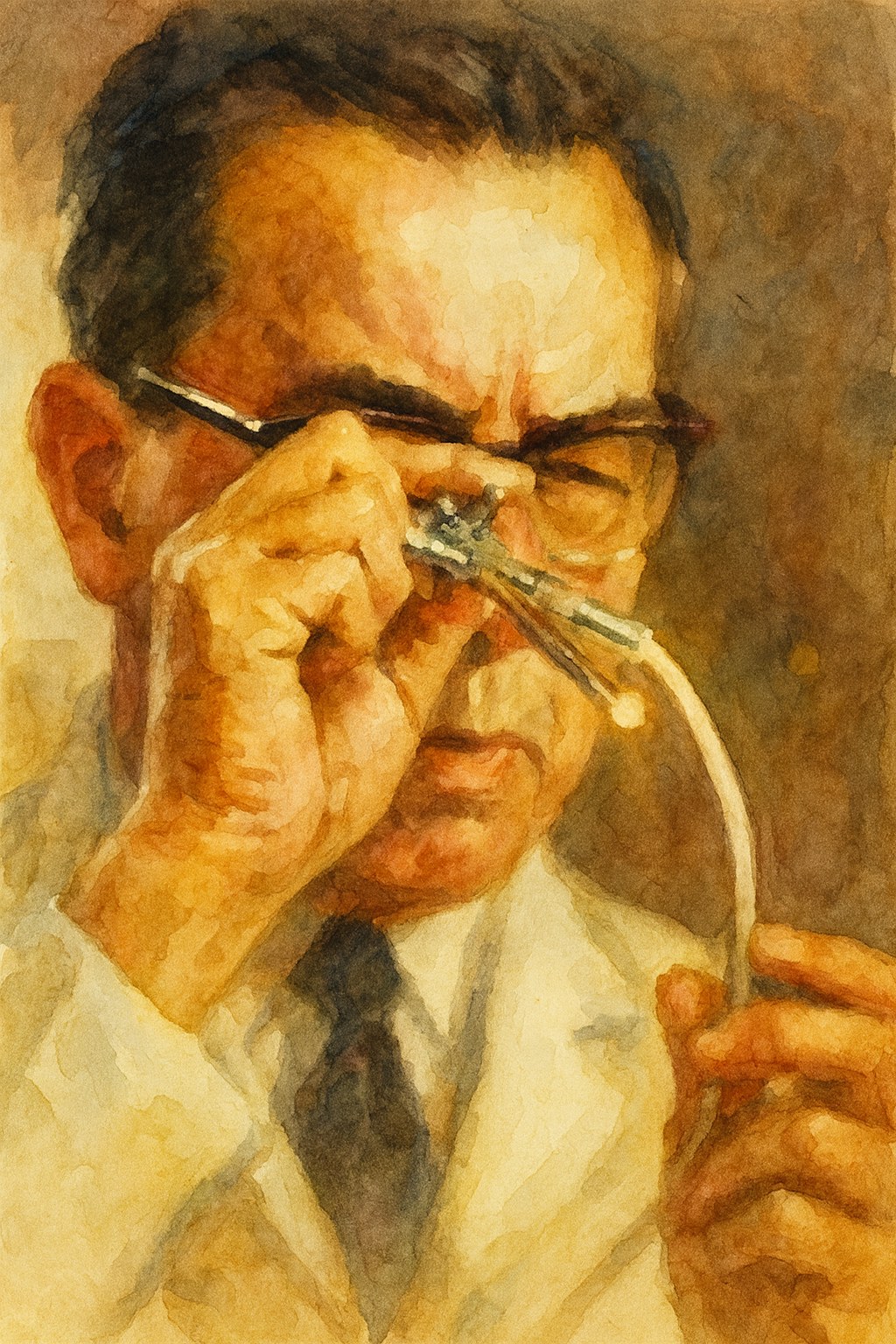
The 1960s brought a revolutionary tool that transformed meniscal treatment, the arthroscope. Japanese orthopedic surgeon Masaki Watanabe, 1911 to 1995, perfected the first practical arthroscopes. He built on earlier work by Dr. Kenji Takagi, who attempted knee endoscopy in Tokyo in 1918, and by Swiss surgeon Dr. Eugen Bircher in the 1920s.
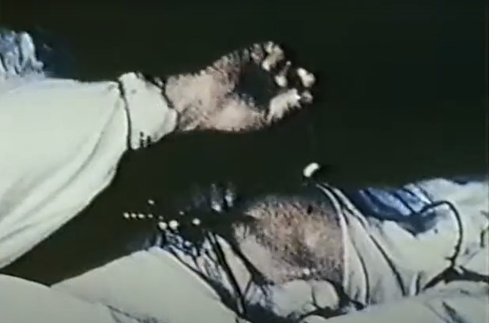
Watanabe developed the No. 21 arthroscope in 1958, featuring a small diameter lens and improved lighting. For the first time, surgeons could see clearly inside the knee joint without opening it widely. On May 4, 1962, Watanabe made history by performing the world’s first arthroscopic meniscectomy, removing a torn meniscal fragment through tiny portals under direct scope view.
Arthroscopic meniscectomy was a radical departure from open arthrotomy. Watanabe went on to publish an Atlas of Arthroscopy with vivid photographs, inspiring others. By the late 1960s, he and colleagues, including Dr. Hideshige Moriya, were refining arthroscopic techniques and instruments.
While arthroscopy was initially used mostly as a diagnostic tool, Watanabe’s 1962 case proved it could be therapeutic. His contributions earned him the title “father of modern arthroscopy.” The knee was no longer a black box that had to be opened widely to be understood. The stage was set for an arthroscopic revolution in the 1970s, drastically reducing the morbidity of meniscus surgery and enabling new procedures like partial meniscectomy and meniscal repair with much less invasive means.
1970s
The Shift Toward Meniscal Preservation
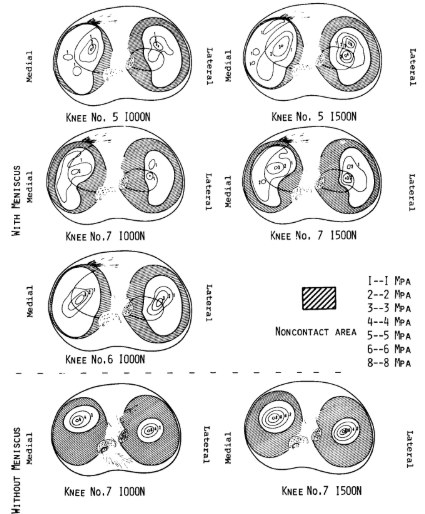
Fukubayashi T, Kurosawa H. The contact area and pressure distribution pattern of the knee. A study of normal and osteoarthritic knee joints. Acta Orthop Scand. 1980;51(6):871-879.
Despite earlier insights, through the 1960s many surgeons still treated meniscus tears with total meniscectomy, often via open surgery. In the 1970s, accumulating evidence accelerated a paradigm shift.
Biomechanical studies, such as Krause et al. in 1976 in Germany, quantified the meniscus’s load bearing role, showing that the menisci transmit a significant portion of knee forces, approximately 50% on the medial side and 70% on the lateral side. Another study by Ahmed and Burke found similar load distribution percentages. These data reinforced that removing the meniscus can greatly increase contact pressure on cartilage, raising the risk of early arthritis.
Long-term clinical follow-ups also confirmed high rates of osteoarthritis after meniscectomy. Consequently, surgeons began advocating partial meniscectomy, trimming only the torn portion and preserving as much meniscus as possible. For a broader audience, this was the central lesson, the meniscus was not disposable padding, but a load sharing structure that protected the joint.
Meniscal preservation, radical in earlier decades, gained traction as arthroscopic techniques made limited resection feasible. For example, British surgeon J. Fairbank’s son, Harold Fairbank, and others in the 1970s promoted leaving stable meniscal remnants intact rather than “whipping out” the entire meniscus.
Toru Fukubayashi published biomechanical data demonstrating that removal of one meniscus shifted the load to the remaining intact meniscus, simply and powerfully providing evidence in support of meniscus preservation. By the early 1980s, the tide had turned. Partial meniscectomy, especially arthroscopic partial meniscectomy, became the new gold standard, replacing total meniscectomy for many tears. This shift marked one of the most significant changes in orthopedic sports medicine.
“In anatomical terms, there are some differences in the medial and lateral menisci. The lateral meniscus is a round C-shape compared with the medial one, which has a lazy C-shape. Moreover, the lateral meniscus has a free periphery not attached to the capsule or tibia in its posterior half and therefore has more mobility than the medial meniscus, which is fixed along its entire periphery by the meniscofemoral and meniscotibial ligaments.
From the present study, it was found that the lateral meniscus occupied a greater percentage area in the lateral compartment than did the medial meniscus in the medial compartment, and the difference is significant (P < 0.01). Accordingly, it may be said that the lateral meniscus contributes more to weight-bearing in the lateral compartment than does the medial meniscus in the medial compartment. In contrast, the medial meniscus plays a more important role in joint stability than does the lateral meniscus, judging from both its anatomy and our results.”

1982
Vascular Zones and the Science of Meniscal Healing
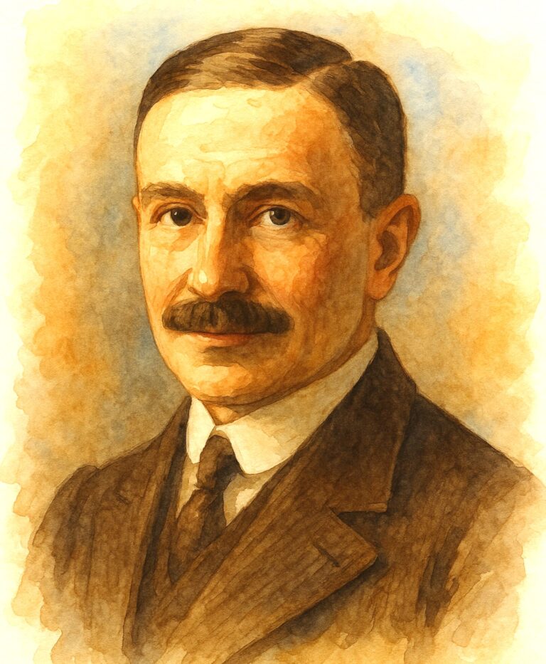
Arnoczky SP, Warren RF. The microvasculature of the meniscus and its clinical significance. Clin Orthop Relat Res. 1982;(163):94-101.
A milestone discovery in 1982 helped explain why some meniscus tears heal while others do not. Veterinary surgeon Steven P. Arnoczky and orthopedic surgeon Russell F. Warren at Hospital for Special Surgery published their classic study on the microvascular anatomy of the meniscus. Repair was no longer just optimism. It now had anatomy behind it.
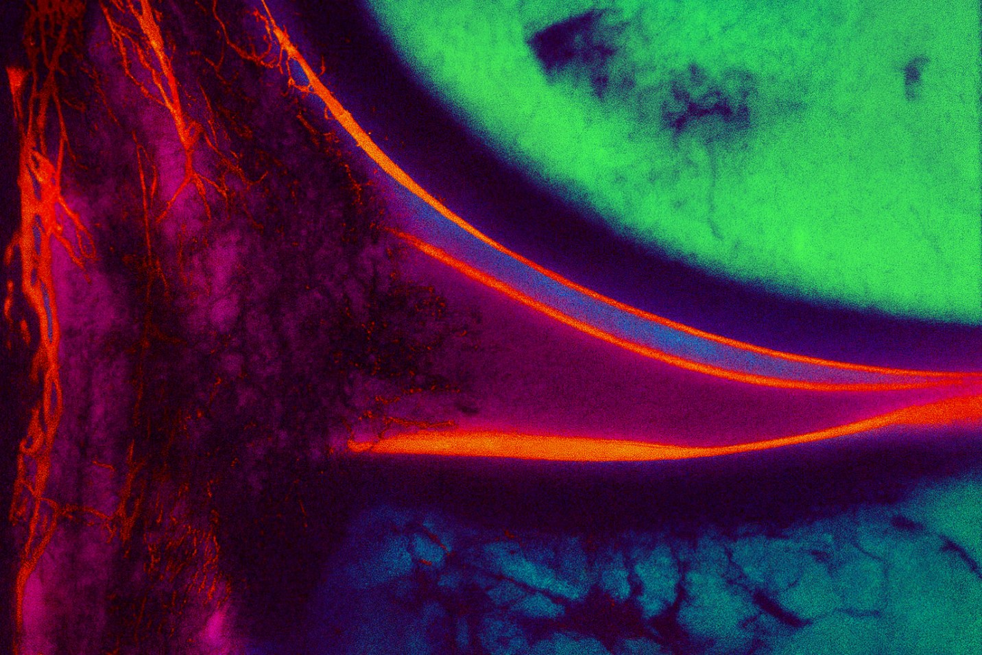
Examining human cadaver menisci with special tissue clearing techniques, they showed that blood vessels penetrate only the outer 10 to 25% of the meniscus width, an area now known as the “red zone.” The inner “white zone” receives nutrition only from synovial fluid. They also observed a well vascularized synovial fringe around the meniscus periphery and good blood supply in the meniscal horn attachments.
Arnoczky and Warren’s work established the fundamental concept of red-red, red-white, and white-white zones to classify tear locations by healing potential. A tear in the red-red zone, peripheral, has a high chance of healing, especially if repaired. A red-white zone tear, at the junction, has intermediate potential. A white-white, central tear, has poor healing capacity.
Arnoczky and Warren’s paper, widely cited, provided the anatomic rationale for meniscus repair. Tears in vascular regions have the best chance to heal. The study directly informed surgical algorithms, encouraging attempts to suture tears in the outer third while acknowledging that central tears would often require resection. Indeed, Arnoczky later recounted that this “lucky break” finding came as Dr. DeHaven was beginning to fix peripheral tears, and it spurred many others to try repair instead of resection for red zone tears.
“These observations have led to increasing efforts by orthopedic surgeons to repair certain meniscal lesions. The results of this study would suggest that lesions of the peripheral attachment of the menisci, as well as those involving the anterior or posterior horn attachments, have access to a vascular supply which may be adequate enough to support a healing response. An exception would be lesions in the posterolateral aspect of the lateral meniscus where the blood supply is tenuous.”
1980s and 1990s
MRI Emerges as a Turning Point in Diagnosis
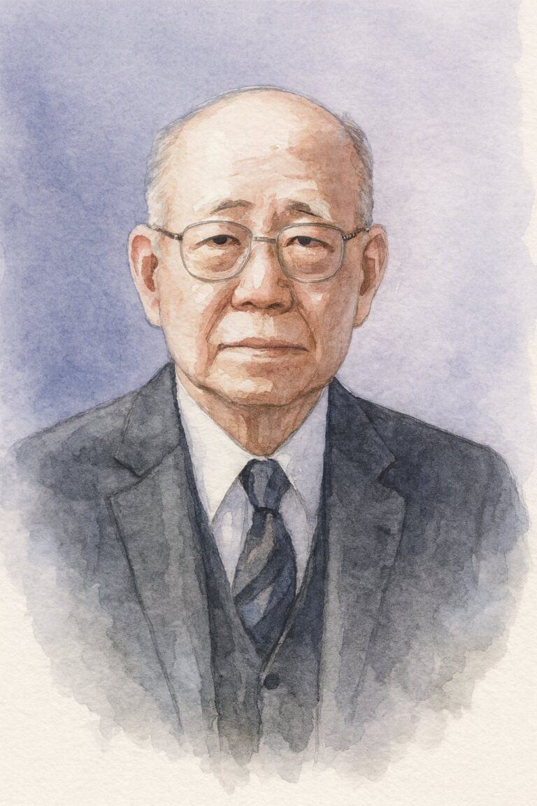
Reicher MA, Bassett LW, Gold RH, Lufkin RB. Magnetic resonance imaging of the knee: patterns of meniscal pathology. Radiology. 1985;155(3):647-651.
After arthroscopy, preservation, and the science of healing had changed how surgeons thought about the meniscus, magnetic resonance imaging, MRI, transformed how they could see it before surgery. Throughout the 1970s and early 1980s, whole body scans were explored by pioneers including John R. Mallard and his team at the University of Aberdeen, Scotland. Different body parts were then isolated, with authors such as Jerry Herman Mink, a musculoskeletal radiologist in Los Angeles, publishing Magnetic Resonance Imaging of the Knee in 1984.
Murray A. Reicher and his group at UCLA published on meniscus pathology seen on MRI in 1985. MRI soon became a preferred, noninvasive way to diagnose meniscal tears. By 1984 to 1988, studies demonstrated MRI’s high accuracy in depicting meniscal pathology, with reported sensitivities around 88 to 98% for tears.
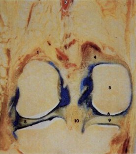
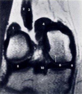
For the first time, radiologists could directly see meniscus tissue and tears on scans, rather than inferring them from arthrogram outlines. MRI could show horizontal cleavage tears, radial tears, and complex degenerative tears in detail. Although early MRI resolution was modest, it quickly replaced routine diagnostic arthroscopy and arthrography in many settings.
Surgeons now had a powerful tool to confirm meniscus diagnoses preoperatively, or even manage some cases nonoperatively if MRI showed a stable tear. By the 1990s, MRI was ubiquitous, an essential part of the meniscal injury workup. MRI greatly improved decision making and helped identify subtle pathology like meniscal cysts or discoid meniscus variants that earlier might have been missed. For patients, the practical meaning was simple, the knee could often be understood before anyone made an incision. It is the cornerstone for the diagnosis of meniscal pathology today.
“The potential roles of MRI of the knee joint include (1) evaluation of a suspected structural abnormality; we are currently pursuing clinical trials to assess the accuracy of MRI in a wide diversity of posttraumatic and nontraumatic structural and/or functional abnormalities; and (2) evaluation and demonstration of dynamic anatomy and physiology; since MRI permits direct visualization without anatomic dissection of living ligaments, tendons, and muscles, it is a unique tool of the in vivo investigation of function and biomechanics. The images also provide a valuable tool for teaching anatomy.”
Conclusion
Together, the advances from this era allowed surgeons to see and understand the meniscus more deeply and accurately. No longer was the diagnosis made solely on clinical acumen. Objective data now came from imaging and direct visualization with arthroscopy.
Arthroscopy allowed surgeons to address meniscal defects more precisely. That precision made partial meniscectomy practical and created a platform for meniscal repair.
The meniscus in the 1980s and 1990s was transformed from a structure often removed to one increasingly seen as worth saving, and increasingly possible to save. By the end of the 1980s, improved imaging, arthroscopic technique, biomechanical insight, and vascular anatomy had laid the foundation for modern meniscal preservation, a warmer, wiser era of care centered on seeing more clearly and removing less.
References
- Annandale T. An operation for displaced semilunar cartilage. Br Med J. 1885;1(1268):779. doi:10.1136/bmj.1.1268.779
- Katzenstein M. Beitrag zur Entstehung und Erkennung der Meniscusverletzungen im Kniegelenk. Berliner Klinische Wochenschrift. 1908;44:228-235.
- King D. The function of semilunar cartilages. J Bone Joint Surg Am. 1936;18(4):1069-1076.
- McMurray TP. The semilunar cartilages. Br J Surg. 1942;29(116):407-414. doi:10.1002/bjs.18002911612
- Fairbank TJ. Knee joint changes after meniscectomy. J Bone Joint Surg Br. 1948;30-B(4):664-670. PMID: 18894618
- O’Donoghue DH. Surgical treatment of fresh injuries to the major ligaments of the knee. J Bone Joint Surg Am. 1950;32(4):721-738.
- Warren RF. Arthroscopic meniscus repair. Arthroscopy. 1985;1(3):170-172.
- DeHaven KE, Merryweather J, Hutton W, et al. Associated tears of the menisci and articular surfaces in acute ruptures of the anterior cruciate ligament. Am J Sports Med. 1980;8(6):503-510.
- Jones RW. Specimen of internal semilunar cartilage as a complete disc. Proc R Soc Med. 1930;23(11):1588-1589. doi:10.1177/003591573002301141
- Arnoczky SP, Warren RF. Microvasculature of the human meniscus. Am J Sports Med. 1982;10(2):90-95. PubMed ID: 7081532. https://pubmed.ncbi.nlm.nih.gov/7081532/
- Pearse EO, Craig DM. Three Jewish Germans as chief surgeons in the Berlin-Friedrichshain Hospital, a historical reminiscence. Zentralbl Chir. 2005;130(5):490-495. PubMed ID: 16220448. https://pubmed.ncbi.nlm.nih.gov/16220448/
- Thomas Annandale: The first meniscus repair. ResearchGate. https://www.researchgate.net/publication/236192165_Thomas_Annandale_The_first_meniscus_repair
- Fairbank TJ. Review of meniscectomy. PMC. https://pmc.ncbi.nlm.nih.gov/articles/PMC3438273/
- Meniscal repair and regeneration: Current strategies and future perspectives. PMC. https://pmc.ncbi.nlm.nih.gov/articles/PMC6128795/
- Biomechanics of the menisci of the knee. ScienceDirect. https://www.sciencedirect.com/science/article/abs/pii/S0268089008000613
- Unhappy triad of the knee: What are the current concepts and opinions? PMC. https://pmc.ncbi.nlm.nih.gov/articles/PMC10251265/
- Contrast arthrography of the knee joint. Radiology. 1967;89(6):1083. https://pubs.rsna.org/doi/10.1148/89.6.1083
- Knee arthroscopy in the era of precision medicine: A comprehensive review. PMC. https://pmc.ncbi.nlm.nih.gov/articles/PMC11537776/
- Giants of orthopaedic surgery: Masaki Watanabe MD. PMC. https://pmc.ncbi.nlm.nih.gov/articles/PMC3705061/
- Meniscal ramp lesions: Rediscovering the past. Knee Surg Sports Traumatol Arthrosc. 2022. https://link.springer.com/article/10.1007/s00167-022-07184-z
- Meniscal tear, presentation, diagnosis and management. Aust Fam Physician. 2012. https://www.racgp.org.au/afp/2012/april/meniscal-tear
- The CT knee arthrogram revisited. PMC. https://pmc.ncbi.nlm.nih.gov/articles/PMC10860528/
- Meniscal transplantation: State of the art. ScienceDirect. https://www.sciencedirect.com/science/article/pii/S2059775421002613
- Meniscus transplantation and anterior cruciate ligament replacement. PubMed. 1990. https://pubmed.ncbi.nlm.nih.gov/2382204/
- The discoid meniscus. PMC. https://pmc.ncbi.nlm.nih.gov/articles/PMC2656711/
- Discoid meniscus. StatPearls. NCBI Bookshelf. https://www.ncbi.nlm.nih.gov/books/NBK470370/
- Discoid meniscus. Orthobullets. https://www.orthobullets.com/knee-and-sports/3007/discoid-meniscus
- Meniscal root tear repair: Why, when and how? PMC. https://pmc.ncbi.nlm.nih.gov/articles/PMC4508558/
- Meniscal root tear. Radiopaedia. https://radiopaedia.org/articles/meniscal-root-tear?lang=us
- Meniscus root tears: What is the hype? Singapore Med J. https://journals.lww.com/smj/fulltext/9900/meniscus_root_tears__what_is_the_hype_.147.aspx
- LaPrade RF. Meniscal root tear classification 2014. https://drrobertlaprademd.com/wp-content/uploads/2015/07/Meniscal-Root-Tear-Classification-2014.pdf