0:00
Introduction
The history of meniscal pathology offers a powerful example of how orthopedic paradigms evolve. In 1897, anatomist Sir Arthur Bland-Sutton dubbed the menisci “functionless remnants of intra-articular leg muscles,” reflecting the dominant belief that the meniscus served no important role. Over the next seven decades, that view was repeatedly challenged and ultimately overturned, giving rise to what we now recognize as essential truth: the meniscus is critical for knee stability, load transmission, and long-term joint health.
That transformation did not unfold in a straight line. Ideas introduced early sometimes fell out of favor, only to be rediscovered decades later as surgical techniques, imaging, and biomechanical science advanced. Progress came through observation, debate, experimentation, and technological change-a century-long process of refining, revising, and occasionally reversing what the field believed it knew.
In this series of articles, we trace the major milestones in the understanding and treatment of meniscal injury, emphasizing the foundational studies, transformative innovations, and key figures that shaped the field. Although many early practices were ultimately abandoned, particularly after the advent of arthroscopy, the insights behind them helped establish the principles that continue to guide modern knee surgery.
The history of the meniscus is not simply a record of changing techniques, but a continuing lesson in how surgical progress depends on reexamining what earlier generations believed they already understood.
The Debate of Meniscal Function in the 1880s – 1969
1883
Thomas Annandale’s First Meniscus Repair
Annandale T. An operation for displaced semilunar cartilage. Br Med J. 1885;1(1268):779. doi:10.1136/bmj.1.1268.779.
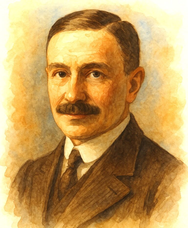
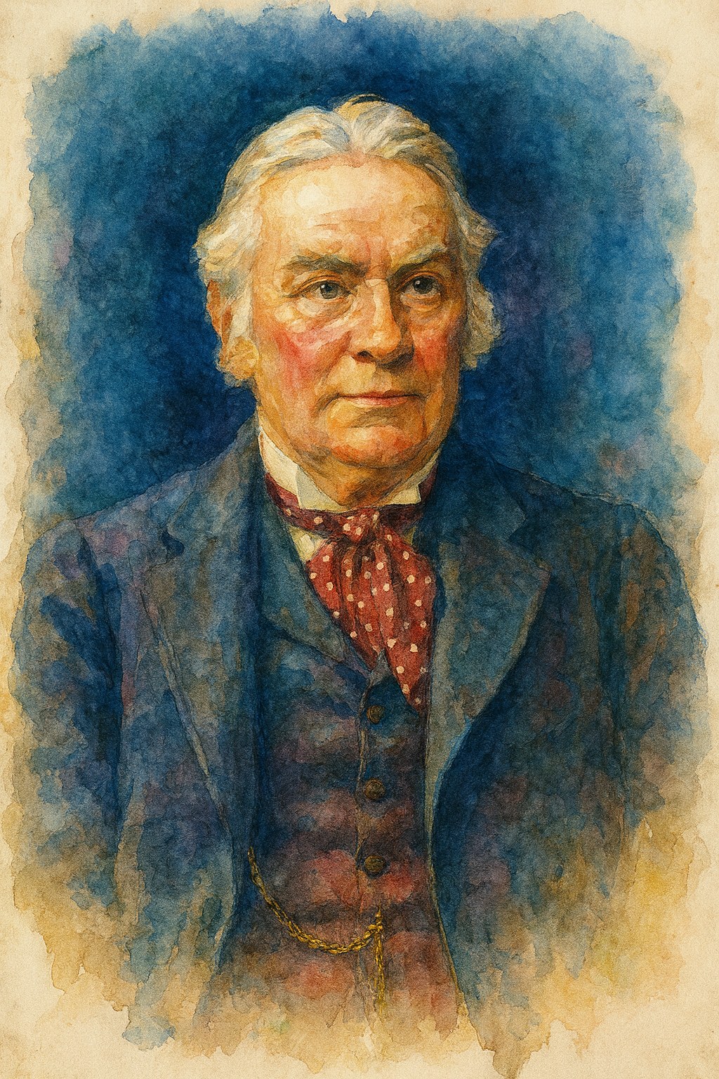
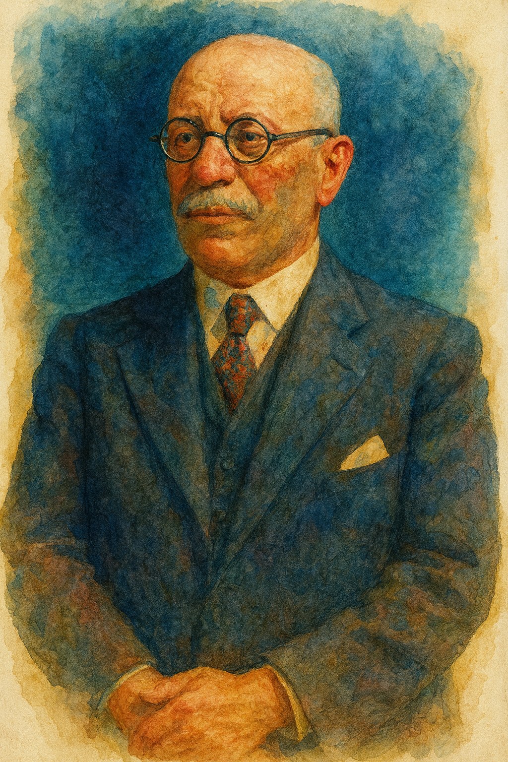
Scottish surgeon Thomas Annandale (1838-1907) is credited with the first recorded meniscus repair. In 1883, he sutured a torn medial meniscus and published the result in 1885. Annandale’s operation, “an operation for displaced semilunar cartilage,” challenged prevailing wisdom by attempting to restore rather than excise the meniscus. His forward-thinking approach did not immediately change practice-the meniscus would still be considered expendable for decades-but it planted the seed for meniscal preservation.
“The pathology of the condition called by that wise old surgeon Hey, of Leeds, “internal derangement of the knee-joint;” by Sir Astley Cooper, “partial luxation of the thigh-bone from the semilunar cartilages;” and, which is now by some authors termed dislocation, or displacement of the semilunar cartilages, has not yet been thoroughly worked out, as few opportunities occur for the dissection of a joint so affected. It is, however, a clinical fact that one of the semilunar cartilages, usually the internal one, does occasionally become loosened from its attachments; and, in consequence, this body is liable to be displaced either forwards or backwards, and so to interfere with the proper movements of the knee-joint.”
“An incision was made along the upper and inner border of the tibia, parallel with the anterior margin of the internal semilunar cartilage; and the few superficial vessels having been secured, the joint was opened. It was then seen that this semilunar cartilage was completely separated from its anterior attachment, and was displaced backwards about half an inch. The anterior edge of this cartilage was now seized by a pair of artery forceps, and it was drawn forwards into its natural position, and held there until three stitches of chromic catgut were passed through it and through the fascia and periosteum covering the margin of the tibia. The forceps were then withdrawn, the cartilage remaining securely stitched in position. The wound in the synovial membrane and soft textures having been closed with catgut stitches, a splint and plaster-of-Paris bandage were applied, so as to keep the joint at rest. The progress of the patient, after the operation, was perfect, the temperature never rising above 99° Fahr. Seven weeks after the operation, the splint and bandages were removed, and gentle movements of the joint practised.”
1908
Moritz Katzenstein Advances Meniscal Surgery
Katzenstein M. Beitrag zur Entstehung und Erkennung der Meniscusverletzungen im Kniegelenk. Berliner Klinische Wochenschrift. 1908;44:228-235.
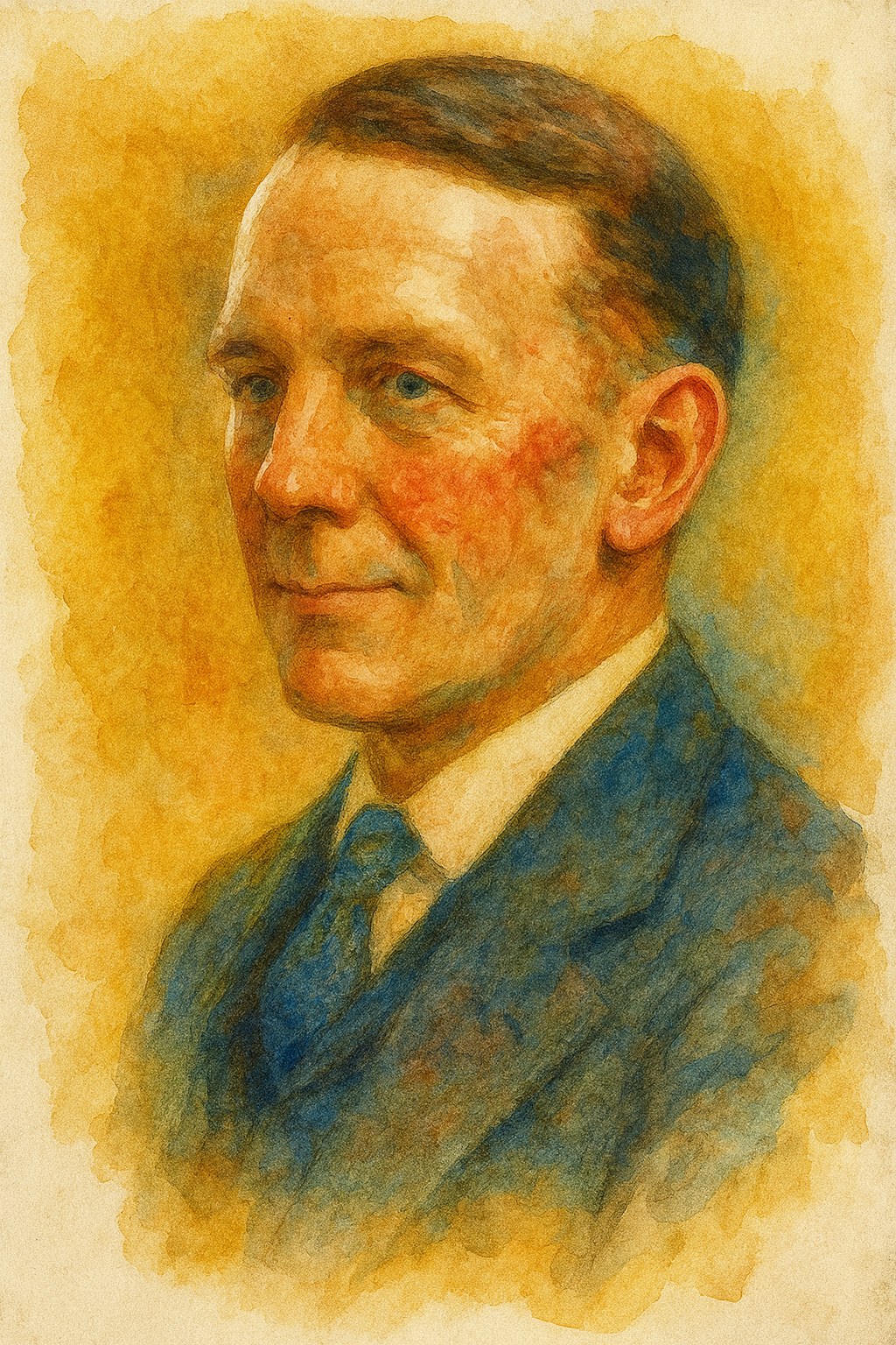
German surgeon Moritz Katzenstein (1872-1932), who served as chief of surgery in Berlin, built on Annandale’s idea. In 1900, Katzenstein performed the first successful meniscal repair in Germany, suturing a torn meniscus back in place. He later published a 1908 case series on meniscus injuries, becoming an advocate for meniscal repair over removal.
Katzenstein even presciently noted that preserving the meniscus could help prevent gonarthrosis-the early term for knee arthritis-in the long term. His pioneering work, largely overlooked at the time, earned him recognition as the “father of meniscus repair.”
“The nature of the injury, the swelling of the joint resulting from the bruise following the trauma, indicated an intra-articular injury . The characteristic entrapment symptoms suggest interposition of the detached joint fragment, and the present pain proves that the injury occurred on the inside of the joint. X-rays performed in most cases reveal the absence of bone injury , and based on the totality of these symptoms, a diagnosis of meniscus injury can be made with some degree of probability.”
“I would strongly recommend suturing the injured meniscus . Not only should the meniscus detached from the anterior or posterior horn and remaining in place be sutured to its undersurface. It is also possible to reposition severely dislocated cartilage and reattach it by suturing. Even if it has shriveled, it can be released from its coiled state through shallow incisions. And even if it cannot be pulled forward all the way around to the edge of the tibia, it is still better than removing it entirely, thereby creating a serious defect. The meniscus, firmly attached to its undersurface, albeit somewhat shortened, perfectly fulfills its function, as my case II shows.”
1930s – 1940s
Physical Examination of the Meniscus and Exploration of its Function
King D. The function of semilunar cartilages. J Bone Joint Surg Am. 1936;18(4):1069-1076. McMurray TP. The semilunar cartilages. Br J Surg. 1942;29(116):407-414. doi:10.1002/bjs.18002911612.
By the 1930s, attitudes began to change. In 1936, American surgeon Dr. Don H. King published a pivotal paper, “The Function of the Semilunar Cartilages,” which provided evidence that the meniscus is not a vestigial structure but rather plays crucial roles in load bearing and joint stability.
King’s experiments, including animal studies, showed that removing the meniscus altered knee mechanics and suggested the meniscus even had some healing capacity at its periphery. This was the first scientific confirmation that the meniscus had important biomechanical functions.
King famously observed, “A torn meniscus can be healed by connective tissue if the tear communicates with the synovial membrane laterally,”- noting that peripheral meniscus tears, which have blood supply from the joint capsule, might repair themselves. His work cracked the door open for meniscal preservation by highlighting the meniscus’s role in knee health.
Around the same time, clinicians also started identifying specific meniscal injury patterns. The term “bucket-handle tear” (a large displaced flap of meniscus) entered the lexicon, explaining cases of the locked knee.
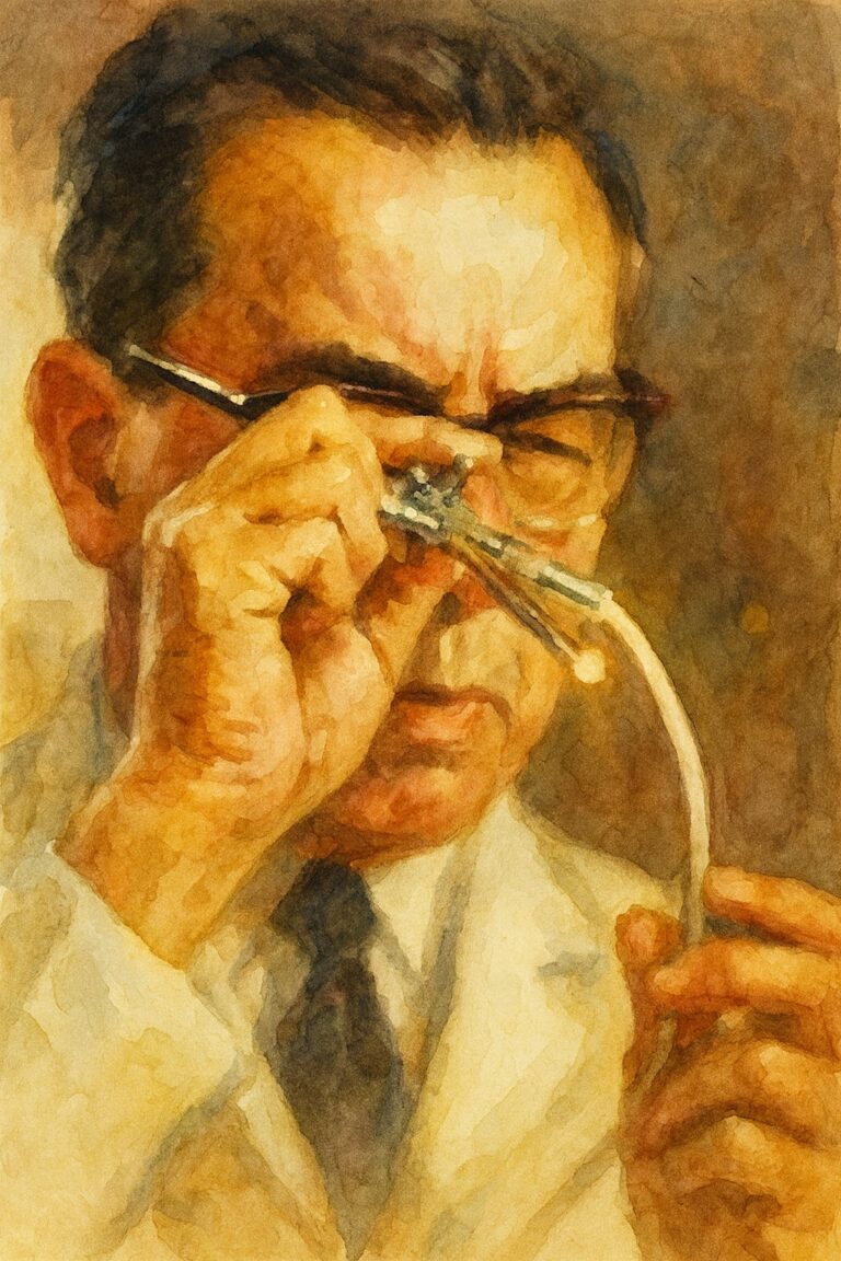
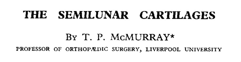
Building on King’s work, clinicians began devising tools to detect meniscal injuries in the clinic. Thomas McMurray (pictured above) described his classic provocative test in 1942, noting that incomplete removal of a torn meniscus often led to persistent symptoms. While McMurray’s test became a staple of diagnosis still used today, his surgical philosophy still favored complete meniscectomy if any fragment remained-reflecting the prevailing instinct to “clean out” the meniscus for a definitive cure.
“In carrying out the manipulation with the patient lying flat, the knee is first fully flexed until the heel approaches the buttock; the foot is then held by grasping the heel and using the forearm as a lever. The knee being now steadied by the surgeon’s other hand, the leg is rotated on the thigh with the knee still in full flexion. During this movement the posterior section of the cartilage is rotated with the head of the tibia, and if the whole cartilage, or any fragment of the posterior section, is loose, this movement produces an appreciable snap in the joint. By external rotation of the leg the internal cartilage is tested, and by internal rotation any abnormality of the posterior part of the external cartilage can be appreciated. By altering the position of flexion of the joint the whole of the posterior segment of the cartilages can be examined from the middle to their posterior attachments. Thus, if the leg is rotated with the knee at right angles the cartilages in their mid-section come under pressure, but, anterior to this point, the pressure exerted on the cartilage is so diminished that accurate examination is impossible.”
“This method of examination is not easy to master; the rotation requires a considerable amount of practice, and the whole procedure must be carried out systematically if success is to be attained. Probably the simplest routine is to bring the leg from its position of acute flexion to a right angle, whilst the foot is retained first in full internal, and then in full external rotation. Any abnormality in the cartilage structure in the area under examination will be discovered during the straightening of the joint.”
1948
Fairbank’s Findings on Meniscectomy Open Eyes to Long-Term Consequences
Fairbank TJ. Knee joint changes after meniscectomy. J Bone Joint Surg Br. 1948;30-B(4):664-670. PMID: 18894618.
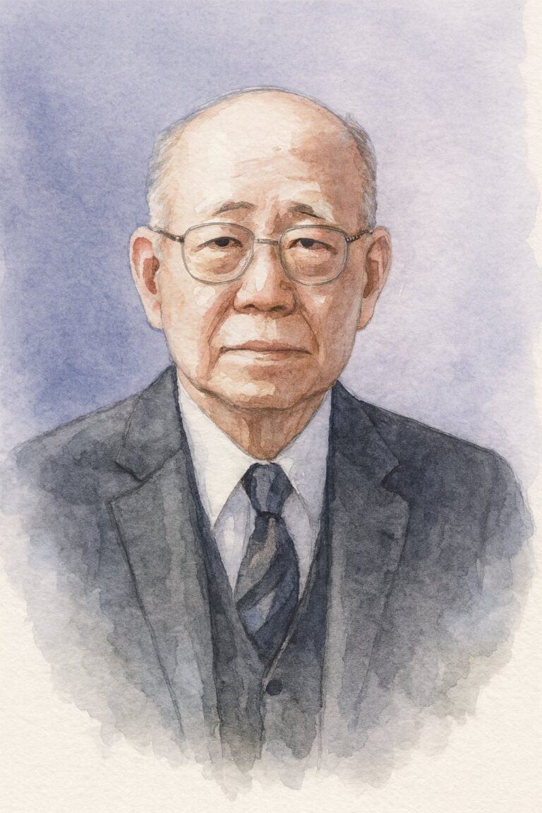
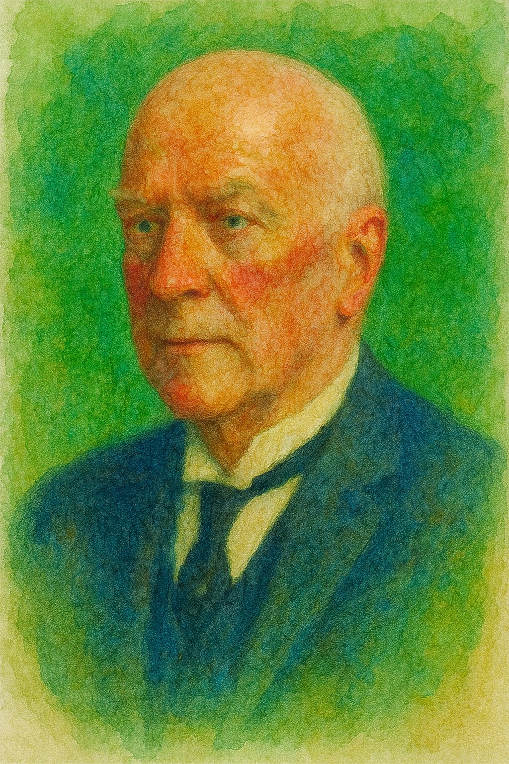
After World War II, advances in radiology brought a crucial insight into view. Thomas “Jack” Fairbank, a British surgeon, reviewed X-rays of patients years after total meniscectomy. In 1948, he reported characteristic post-meniscectomy radiographic changes-joint space narrowing, ridge formation, and flattening of the femoral condyle-now collectively known as “Fairbank’s changes.” Fairbank concluded that losing the cushioning function of the meniscus predisposed the knee to degeneration.
This landmark observation provided clinical evidence that the meniscus is essential for long-term joint health. It reinforced King’s biomechanical findings and underscored the cost of the then-standard total meniscectomy. Fairbank’s work slowly shifted surgical thinking toward meniscal preservation, though change would be gradual.
His name remains eponymous with the radiographic findings seen after meniscectomy. With the shift toward meniscus-preserving procedures, these changes have become less common-a testament to how much Fairbank’s early warning shaped subsequent practice.
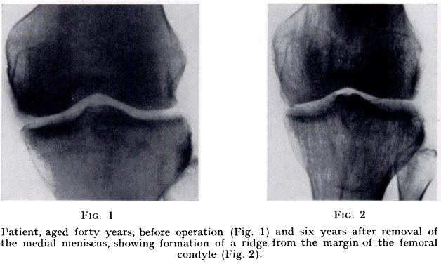
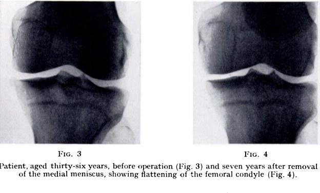

“Changes in the knee joint after meniscectomy include ridge formation, narrowing of the joint space, and flattening of the femoral condyle. Investigations suggest that these changes are due to loss of the weight-bearing function of the meniscus. Meniscectomy is not wholly innocuous; it interferes, at least temporarily, with the mechanics of the joint. It seems likely that narrowing of the joint space will predispose to early degenerative changes, but a connection between these appearances and later osteoarthritis is not yet established and is too indefinite to justify clinical deductions.”

1950
O’Donoghue’s “Unhappy Triad” and Associated Injuries
O’Donoghue DH. Surgical treatment of fresh injuries to the major ligaments of the knee. J Bone Joint Surg Am. 1950;32(4):721-738.
King, McMurray, and Fairbank each advanced a different piece of the meniscal puzzle-its function, its diagnosis, and the cost of removing it. What remained was an understanding of how meniscal tears fit within the broader context of knee injury. That insight came from Dr. Don O’Donoghue.
In 1950, American sports surgeon Don O’Donoghue highlighted the frequent association between meniscus tears and ligament injuries in a case series with detailed illustration of both the injury and treatment. He coined the term “unhappy triad”-also called the O’Donoghue triad-to describe the combined injury of the ACL, MCL, and medial meniscus in athletes, often resulting from a single twisting trauma such as a football tackle.
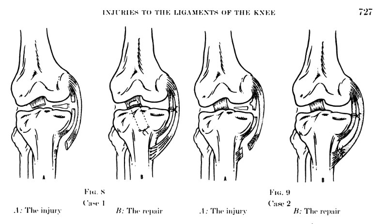
This concept was influential in showing that a meniscal tear often does not occur in isolation. Meniscal pathology is linked with knee instability. For decades, the unhappy triad guided comprehensive treatment of knee injuries. It also spurred research into the role of the meniscus in knee stability: surgeons realized that an intact meniscus helps resist abnormal motion in an ACL-deficient knee, especially the medial meniscus.
He advocated for early repair for all ligaments rather than delay and later reconstruction. Thus, meniscal tears came to be seen as part of a larger injury complex, not just “loose pieces” to remove.
“I must say that in the course of the ten years covered by this report, we have operated upon some twenty patients. This is not a large number when you consider the great many knees that are ordinarily seen in the course of a year. I think this group of athletes is a unique group. They are eighteen to twenty years of age, and at the prime of life; they are in wonderful physical condition, or they would not be athletes. I think our goal here should be complete, 100 per cent recovery. I think we should not willingly consign these patients to a life of relative inactivity. I do not mean to imply that Dr. Bosworth suggests that either, but I want to emphasize that too many times we are inclined to temporize with these cases. The result is that the time for favorable operative results has passed, and we are faced then with a reconstruction, rather than a repair, which is an altogether different problem.”
Conclusion: A Shift in Perspective
By 1969, the meniscus was no longer easy to dismiss as expendable tissue. Early repair attempts by Annandale and Katzenstein, King’s work on function and healing, Fairbank’s warning about degeneration, and O’Donoghue’s view of meniscal tears within larger knee injuries all pointed in the same direction. The meniscus mattered.
Still, understanding came before the tools to act on it.
Surgeons were beginning to see the value of preservation, but open surgery and limited visualization kept meniscectomy at the center of practice. Part I ends in that in-between space-where the importance of the meniscus had become clear, but the modern means to protect it were just beginning to arrive.
The next chapter would bring arthroscopy, better imaging, and a renewed opportunity to treat the meniscus not as something to remove, but as something worth saving.
References
- Annandale T. An operation for displaced semilunar cartilage. Br Med J. 1885;1(1268):779. doi:10.1136/bmj.1.1268.779
- Katzenstein M. Beitrag zur Entstehung und Erkennung der Meniscusverletzungen im Kniegelenk. Berliner Klinische Wochenschrift. 1908;44:228-235.
- King D. The function of semilunar cartilages. J Bone Joint Surg Am. 1936;18(4):1069-1076.
- McMurray TP. The semilunar cartilages. Br J Surg. 1942;29(116):407-414. doi:10.1002/bjs.18002911612
- Fairbank TJ. Knee joint changes after meniscectomy. J Bone Joint Surg Br. 1948;30-B(4):664-670. PMID: 18894618
- O’Donoghue DH. Surgical treatment of fresh injuries to the major ligaments of the knee. J Bone Joint Surg Am. 1950;32(4):721-738.
- Warren RF. Arthroscopic meniscus repair. Arthroscopy. 1985;1(3):170-172.
- DeHaven KE, Merryweather J, Hutton W, et al. Associated tears of the menisci and articular surfaces in acute ruptures of the anterior cruciate ligament. Am J Sports Med. 1980;8(6):503-510.
- Jones RW. Specimen of internal semilunar cartilage as a complete disc. Proc R Soc Med. 1930;23(11):1588-1589. doi:10.1177/003591573002301141
- Arnoczky SP, Warren RF. Microvasculature of the human meniscus. Am J Sports Med. 1982;10(2):90-95. PubMed ID: 7081532. https://pubmed.ncbi.nlm.nih.gov/7081532/
- Pearse EO, Craig DM. Three Jewish Germans as chief surgeons in the Berlin-Friedrichshain Hospital – a historical reminiscence. Zentralbl Chir. 2005;130(5):490-495. PubMed ID: 16220448. https://pubmed.ncbi.nlm.nih.gov/16220448/
- Thomas Annandale: The first meniscus repair. ResearchGate. https://www.researchgate.net/publication/236192165_Thomas_Annandale_The_first_meniscus_repair
- Fairbank TJ. Review of meniscectomy. PMC. https://pmc.ncbi.nlm.nih.gov/articles/PMC3438273/
- Meniscal repair and regeneration: Current strategies and future perspectives. PMC. https://pmc.ncbi.nlm.nih.gov/articles/PMC6128795/
- Biomechanics of the menisci of the knee. ScienceDirect. https://www.sciencedirect.com/science/article/abs/pii/S0268089008000613
- Unhappy triad of the knee: What are the current concepts and opinions? PMC. https://pmc.ncbi.nlm.nih.gov/articles/PMC10251265/
- Contrast arthrography of the knee joint. Radiology. 1967;89(6):1083. https://pubs.rsna.org/doi/10.1148/89.6.1083
- Knee arthroscopy in the era of precision medicine: A comprehensive review. PMC. https://pmc.ncbi.nlm.nih.gov/articles/PMC11537776/
- Giants of orthopaedic surgery: Masaki Watanabe MD. PMC. https://pmc.ncbi.nlm.nih.gov/articles/PMC3705061/
- Meniscal ramp lesions: Rediscovering the past. Knee Surg Sports Traumatol Arthrosc. 2022. https://link.springer.com/article/10.1007/s00167-022-07184-z
- Meniscal tear – presentation, diagnosis and management. Aust Fam Physician. 2012. https://www.racgp.org.au/afp/2012/april/meniscal-tear
- The CT knee arthrogram revisited. PMC. https://pmc.ncbi.nlm.nih.gov/articles/PMC10860528/
- Meniscal transplantation: State of the art. ScienceDirect. https://www.sciencedirect.com/science/article/pii/S2059775421002613
- Meniscus transplantation and anterior cruciate ligament replacement. PubMed. 1990. https://pubmed.ncbi.nlm.nih.gov/2382204/
- The discoid meniscus. PMC. https://pmc.ncbi.nlm.nih.gov/articles/PMC2656711/
- Discoid meniscus. StatPearls. NCBI Bookshelf. https://www.ncbi.nlm.nih.gov/books/NBK470370/
- Discoid meniscus. Orthobullets. https://www.orthobullets.com/knee-and-sports/3007/discoid-meniscus
- Meniscal root tear repair: Why, when and how? PMC. https://pmc.ncbi.nlm.nih.gov/articles/PMC4508558/
- Meniscal root tear. Radiopaedia. https://radiopaedia.org/articles/meniscal-root-tear?lang=us
- Meniscus root tears: What is the hype? Singapore Med J. https://journals.lww.com/smj/fulltext/9900/meniscus_root_tears__what_is_the_hype_.147.aspx
- LaPrade RF. Meniscal root tear classification 2014. https://drrobertlaprademd.com/wp-content/uploads/2015/07/Meniscal-Root-Tear-Classification-2014.pdf