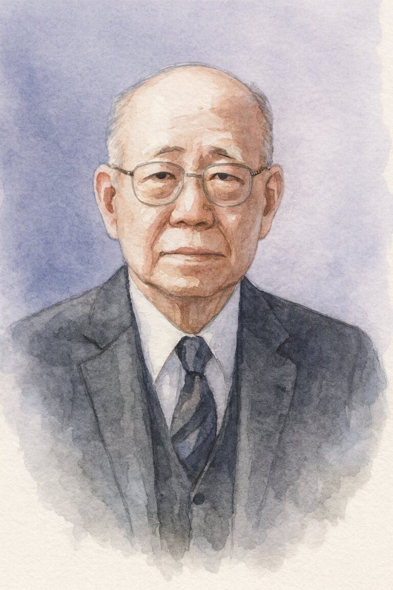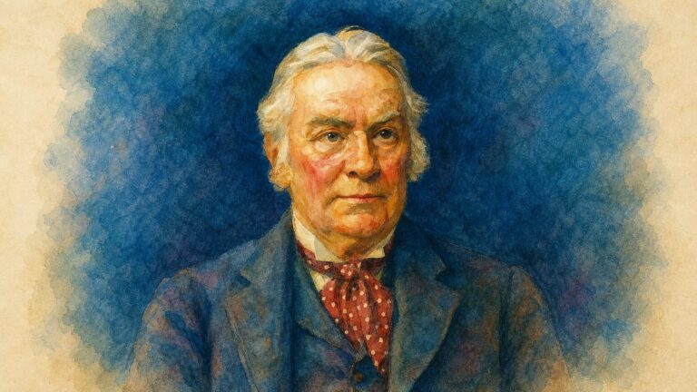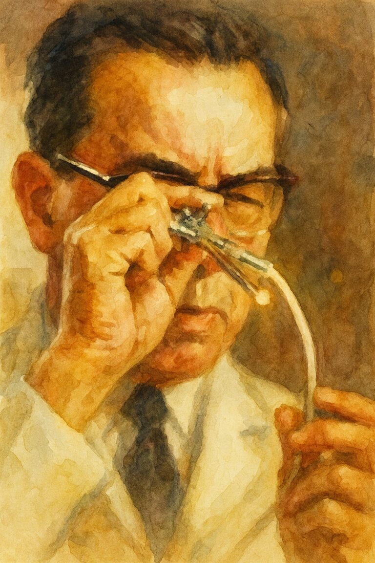
0:00
Shoulder instability has long been a therapeutic dilemma. The anatomic lesions responsible were already well recognized in the early 20th century, and from these discrete lesions on the glenoid and humeral side, countless treatments emerged. Over time, many of these fell out of favor, while a few key techniques were refined and endured. As understanding of shoulder anatomy and biomechanics deepened, indications for treatment also became more clearly defined.
Nearly every aspect of the shoulder instability spectrum carries an eponym, a reflection of the history shaped by dozens of influential and recognizable pioneers. Here, we provide summaries and original works from many of these authors. While some of their key contributions remain cornerstones, others—once highly cited and celebrated in their time—did not stand the test of history.
Foundational Years of Shoulder Instability 1900 – 1969
1906
Georg Perthes, An Operation for Habitual Subluxation of the Shoulder
Perthes G. Über Operationen bei habitueller Schulterluxation. Dtsch Z Chir. 1906;85:199-227. doi:10.1007/BF02894989
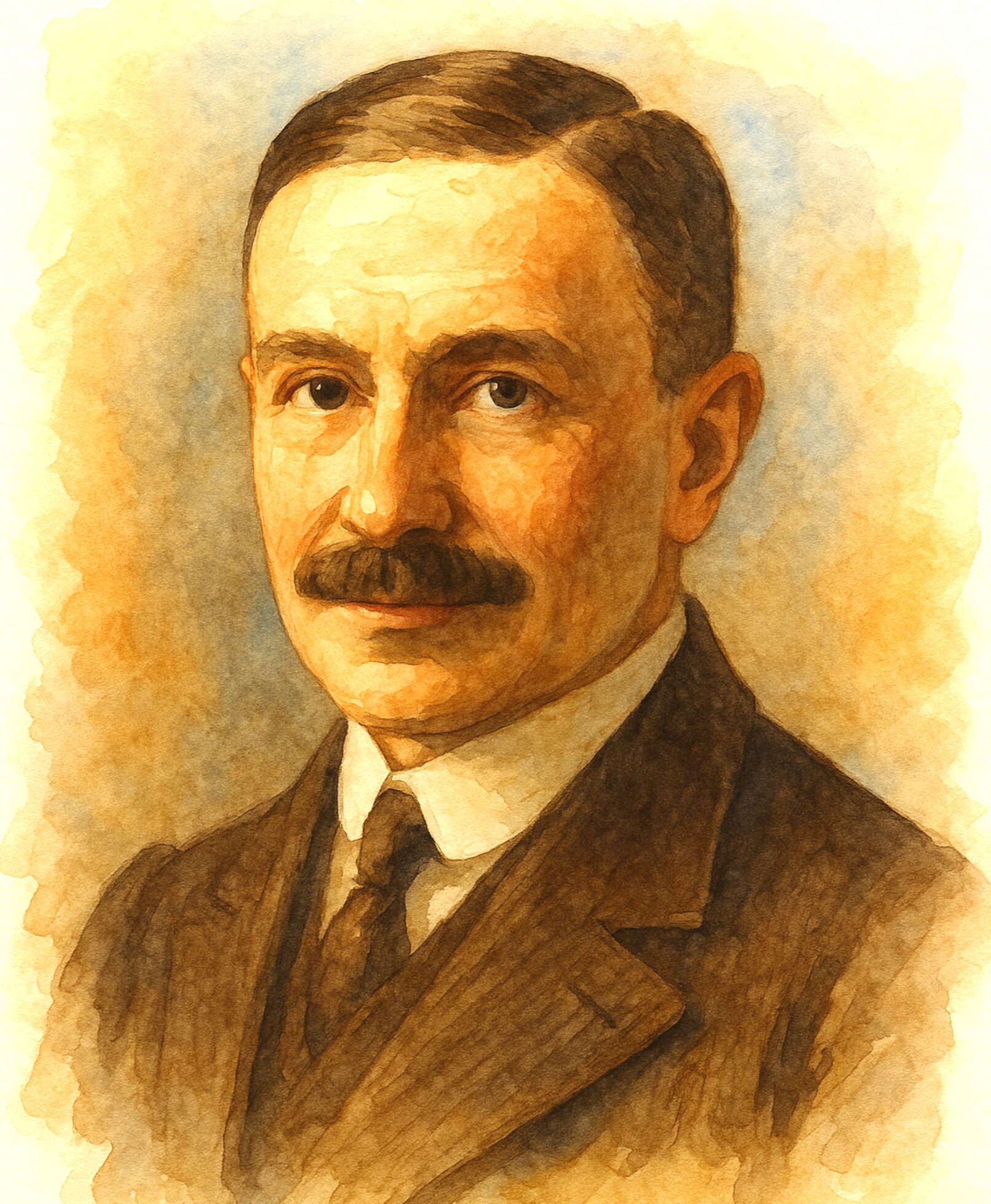
Georg Clemens Perthes (1869–1927) was a German orthopedic surgeon best known for describing the pediatric hip disorder that now bears his name, Legg–Calvé–Perthes disease. Born in Moers, Germany, he trained in Bonn, Berlin, and Leipzig before becoming director of the Orthopedic Clinic in Tübingen in 1907. Perthes was an early adopter of radiology in orthopedics and published influential works on bone and joint tuberculosis, scoliosis, shoulder instability, and hip pathology.
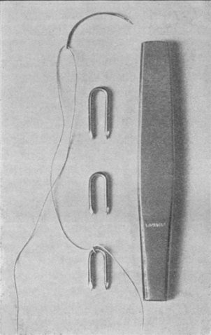
Georg Perthes recognized that recurrent shoulder dislocations were due to detachment of the anterior labrum. He also differentiated labral from rotator cuff pathology. He reported stabilizing the labrum by stapling it to the bone. This was the first reported surgical fixation for shoulder instability.
His legacy endures through his contributions to pediatric orthopedics and his role in advancing modern orthopedic practice.
“The operative treatment of recurrent shoulder subluxations is still not concluded chapter in surgery…Of greater significance it would seem to me, is in a certain group of cases the tearing of the muscles inserting on the major tubercle, and in another group, the tearing of the glenoid labrum on the inner glenoid fossa. The consideration of these pathological changes led me to reconstruct the lost insertion point of the muscles on the major tubercle in two cases, in a further case to reattach the labrum to the glenoid, whilst in a fourth only the reduction and reinforcement of the enlarged and loosened capsule was performed”
1923 & 1938
Arthur S. Blundell Bankart, The Original Open Bankart Repair
Bankart AS. Recurrent or habitual dislocation of the shoulder-joint. Br Med J. 1923;2(3285):1132-1133. doi:10.1136/bmj.2.3285.1132
Bankart AS. The pathology and treatment of recurrent dislocation of the shoulder-joint. Br J Surg. 1938;26(101):23-29. doi:10.1002/bjs.18002610104
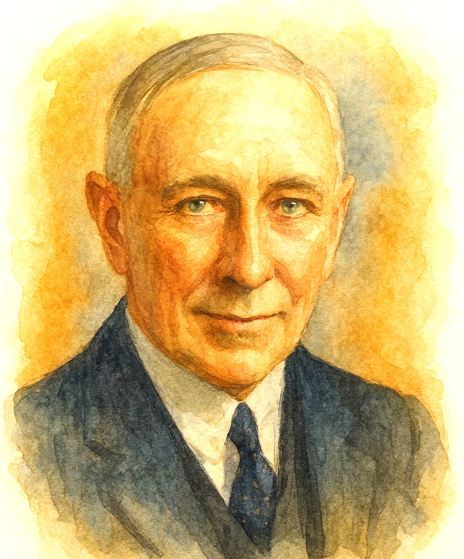
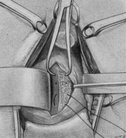
Arthur Sidney Blundell Bankart (1879–1951) was a pioneering British orthopedic surgeon. In 1923 in London he identified the “essential lesion” of recurrent anterior instability as an avulsion of the anteroinferior labrum (later called the Bankart lesion). Bankart pioneered an open repair in which he reattached the torn labrum and capsule to the glenoid rim with sutures, restoring stability without undue loss of motion.
His seminal 1938 paper in the British Journal of Surgery detailed the pathology and treatment of recurrent shoulder dislocation, laying the foundation for modern anatomic repairs. This concept — that repairing the labrum and inferior glenohumeral ligament could cure instability — was transformational in shoulder surgery.
In addition to his influential shoulder work, he contributed to spinal surgery, pain relief through cordotomy, and authored Manipulative Surgery. A founder of both the British Orthopaedic Society and SICOT, Bankart’s legacy endures through his eponymous lesion and operative technique that remain central to shoulder surgery today.
“In my cases the joint defect has been repaired by interrupted sutures of silkworm gut passed between the free edge of the capsule and the glenoid ligament. It is well to freshen the bone on the neck of the scapula, so that the glenoid ligament may adhere to it. Having repaired the joint defect, the divided subscapularis tendon is reunited, the detached portion of the coracoid process is sutured in place, and the wound is closed. After the operation the arm is kept at rest for four weeks, and then active and passive movements are begun and persisted in until the movements of the joint are normal.”
1940
Harold Hill and Maurice Sachs, the Hill–Sachs Lesion of the Humerus
Hill HA, Sachs MD. The grooved defect of the humeral head: a frequently unrecognized complication of dislocations of the shoulder joint. Radiology. 1940;35(6):690-700. doi:10.1148/35.6.690
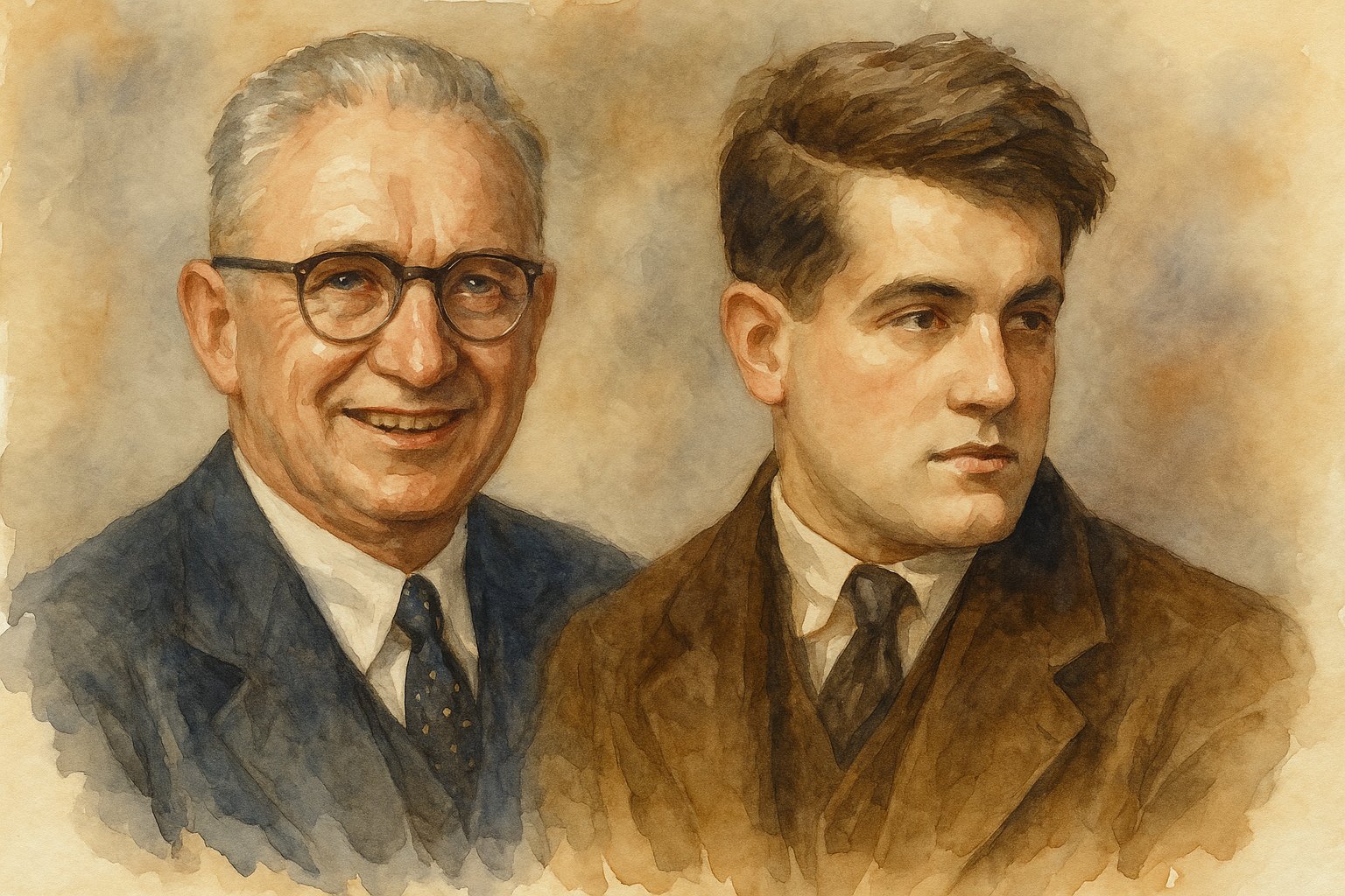
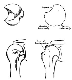
Harold Arthur Hill (1901–1973), depicted left, and Maurice David Sachs (1909–1987), depicted right, were American radiologists whose 1940 landmark study of 119 shoulder dislocations defined the Hill-Sachs lesion, a posterolateral humeral head impaction fracture characteristic of anterior instability. This highly cited work illustrated that bony defects (a “divot” in the humeral head) often accompany dislocations, contributing to instability and guiding later treatment strategies. The recognition of this lesion’s contribution to instability eventually led to the track concept. The lesions are still universally denoted by the combination of their names.
Hill, trained at the University of California, built a private radiology practice in San Francisco and served as a U.S. Navy Reserve captain during World War II, while Sachs, educated at Long Island University and the University of Bern, became professor of radiology at Case Western Reserve University and the founding chair of radiology at Cleveland’s Forest City Hospital, where he advanced both equitable healthcare and early diagnostic imaging. Together, their collaboration cemented a pivotal concept in shoulder pathology and left a lasting legacy in both orthopedics and radiology.
“Compression fractures of the humeral head, unless quite large, will frequently be overlooked unless the x-ray studies are made with the possibility of such in mind. A stereoscopic anteroposterior examination with the arm adducted and in external rotation is not adequate to demonstrate the defect in many instances. An anteroposterior projection made with the arm in marked internal rotation is essential, and in a small percentage of cases the defect will be disclosed only by means of a tangential view of the posterolateral aspect of the humeral head. With marked internal rotation, the posterolateral aspect of the head is viewed in profile so that the length and depth of the compressed area may be evaluated (Fig. 2). The tangential view is necessary to measure the width; this view is obtained by placing the film on the top of the shoulder while the tube is lateral to and below the elbow, the arm being internally rotated.”
1943 & 1948
Capsular Shift Procedures are Introduced
Osmond-Clarke H. Habitual dislocation of the shoulder: the Putti-Platt operation. J Bone Joint Surg Br. 1948;30B(1):19-25.
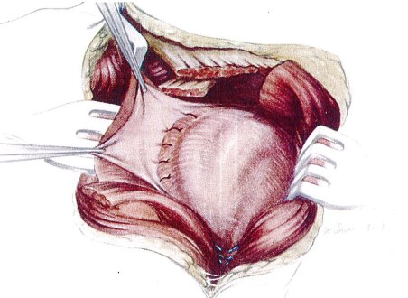
When Bankart’s suturing technique proved technically challenging for some, surgeons explored simpler soft-tissue operations. The Magnuson–Stack procedure (1943) involved shifting the subscapularis tendon insertion laterally to tighten the anterior capsule. The Putti–Platt procedure (described by Oscar Putti and Walton Platt, popularized in 1948) involved splitting and overlapping the anterior capsule and subscapularis to prevent dislocation. These procedures were widely used mid-century to stabilize traumatic anterior instability, though at the cost of reduced external rotation. Later studies by Hawkins and others noted that such non-anatomic tightening could lead to arthritis decades after repair and these techniques fell out of favor.
The Putti-Platt procedure, first performed in 1925, is an anterior stabilization technique for recurrent shoulder dislocation that reinforces the capsule by overlapping and shortening the subscapularis tendon. Through a deltopectoral approach, the subscapularis is divided; its distal stump is sutured to the anterior glenoid rim or capsule, while the proximal stump is attached to the anterior capsule, producing a double layer of restraint. This creates a strong anterior buttress against redislocation, though often at the cost of reduced external rotation.
“During these ten years, however, some surgeons realised that there is not always a gross lesion of the gleno-labral margin. As Platt says in a personal communication: “I evolved my present technique after the first few experiences of carrying out the Bankart procedure as described by him originally. I soon found there was no single and constant ‘Bankartian’ lesion capable of being repaired by a standard procedure. It therefore occurred to me to make sure by stitching the distal end of the divided subscapularis tendon to the cartilaginous remains of the glenoid margin. This provided a primary barrier to redislocation of the head forwards and inwards under the subscapularis. It then appeared logical to stitch the proximal divided end of the subscapularis to the anterior capsule—thus producing an overlap and shortening of the tendon.” This was the genesis of the operation which I have called the Putti-Platt capsulorrhaphy. The first operation was performed at Ancoats Hospital, Manchester, by Platt on November 13, 1925.”
1952
Harrison McLaughlin addresses the Challenge of Posterior Dislocations:
McLaughlin HL. Posterior dislocation of the shoulder. J Bone Joint Surg Am. 1952;24(3):584-590.
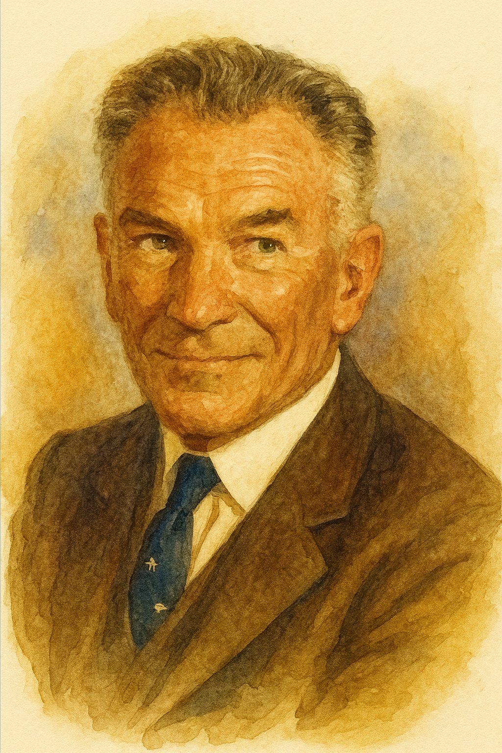
Harrison Lloyd McLaughlin, M.D. (1901–1983), a leader of the Columbia Shoulder Service, was among the earliest pioneers in the study and surgical management of rotator cuff pathology. Beginning his career on the Columbia Fracture Service in 1935, he published influential studies in the 1940s–1950s that emphasized individualized tendon repair and introduced the concept of impingement between the rotator cuff and acromion.
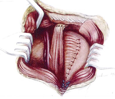
McLaughlin is also remembered for his contributions to the treatment of shoulder instability. He reported on the treatment of locked posterior shoulder dislocations, and later described transferring the subscapularis tendon into the reverse Hill–Sachs defect in the humeral head to prevent recurrence. This operative procedure later bore his name, and his landmark JBJS paper laid the foundation for subsequent leaders in shoulder surgery.
“Posterior dislocation of the humerus is a diagnostic trap. Clinical and roentgenographic evidence of this lesion is always present, but usually escapes notice unless reduced. Recent lesions, recognized and treated at once, respond rapidly and well to simple measures. Recurrent or unreduced lesions require operative treatment. Irreparable damage to the glenohumeral articulation does not always require arthrodesis. The most common actual and serious potential complication of posterior dislocation of the humerus is the failure to arrive at the correct diagnosis prior to commencing treatment. Some failures of surgical treatment for habitual anterior dislocation of the humeral head result from the fact that the recurrent episodes were in reality posterior subluxations of the humerus.”
1954
Michel Latarjet and his Enduring Procedure
Latarjet M. Treatment of recurrent dislocation of the shoulder. Lyon Chir. 1954;49(8):994-997. PMID:13234709
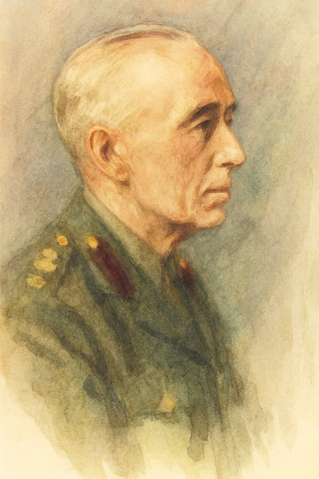
Michel Latarjet (1913–1999) was a French anatomist and surgeon from Lyon. Son of André Latarjet, a renowned anatomist, he carried forward his father’s legacy while developing his own surgical innovations. In 1954 he introduced a revolutionary bony reconstruction in 1954 to address instability with bone loss. The Latarjet procedure involved transferring the coracoid process with attached tendons to the anterior glenoid rim to act as a bone block and muscular sling. Latarjet’s concept provided a two-fold stabilizing effect (increasing the bony depth of the glenoid and adding a dynamic sling via the conjoint tendon) and has proven enduringly influential in treating chronic or bone-deficient shoulder instability.
Beyond orthopedics, Michel was a multifaceted figure—an accomplished sportsman, musician, and academic leader—who revised and expanded the classic Testut-Latarjet anatomy text, served as president of the Société de Chirurgie de Lyon, and contributed extensively to thoracic and musculoskeletal surgery. His eclectic life and enduring surgical contribution remain hallmarks of 20th-century French medicine.
“A smile may come from such a small number. But sometimes the beginnings of an experience are fruitful. When one operates for the first time on recurrent dislocation of the shoulder, it is difficult to decide which technique or procedure to adopt, especially when the patient is young and athletic and has already experienced repeated, disabling relapses.
As Mr. Guilleminet rightly emphasized, important series of operations with sufficiently long follow-up are rare and do not exist in sufficient numbers to leave an impression of complete certainty. For my part, I knew of Albert Trillat’s development of this surgery, and I decided to perform it myself. The lesions were identical in all four patients: the glenoid rim was missing under the coracoid, without fracture of the anterior glenoid. Three other operations carried out in the same area had shown the same lesions: absence of the anterior glenoid rim, with avulsion of the scapulo-subscapular muscles, and up to, if not beyond, the base of the coracoid.
The view of these lesions seems to me to demonstrate that shoulder recurrence does not depend on the gleno-coracoid passage, but on the condition of the anterior glenoid rim and the capsulo-muscular system attached to it, which can be detached, torn, or avulsed. Several authors, Hibbert, Cave, Wirth, Macaulay, have already pointed this out.
The authors who followed this path, in creating these pre-glenoid buttresses, are not the first. The omoplate fragment (scapula), once considered useless and even unfavorable for the placement of the buttress, is the one that is seen, or grafted. Moreover, the coracoid fragment itself, hidden or protruding at the head of the humerus, and on the inside, by the coracoid process, extended by the ligaments and the muscles attached to it.”
1958
Arthur Helfet introduces his Mentor’s Procedure
Helfet AJ. Coracoid transplantation for recurring dislocation of the shoulder. J Bone Joint Surg Br. 1958;40B(2):198-202. doi:10.1302/0301-620X.40B2.198
Arthur Jacob Helfet (1907–1989) was a South African-born orthopaedic surgeon whose career spanned service, leadership, and scholarship across three continents. After early training in Cape Town and Liverpool, he rapidly advanced in orthopaedics. During World War II, he served as an orthopaedic consultant for Allied forces in Africa and published influential work on endemic disease and orthopaedic pathology. Following the war, he played a key role in shaping orthopaedic services in South Africa, including helping establish the College of Physicians, Surgeons and Gynaecologists, and later led accident services in Israel, for which he was awarded the State of Israel Liberation Medal. In 1961, Helfet was appointed Professor of Orthopaedic Surgery at Einstein College of Medicine in New York, where he remained until 1972. A prolific writer and two-time Hunterian Professor, his legacy includes foundational work on meniscal derangements and orthopaedic trauma, along with an enduring influence as a teacher and mentor.
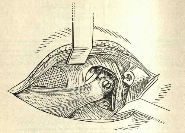
Walter Rowley Bristow (1882–1947), depicted above, was a British orthopaedic surgeon remembered as a pioneer of modern shoulder surgery and as Consulting Orthopaedic Surgeon to the British Army during World War II. Educated at St. Thomas’ Hospital, he served in Gallipoli during World War I before returning to establish the orthopaedic department at St. Thomas’ in 1919. Known as both a gifted clinician and a beloved teacher, he emphasized patient-centered care, often remarking that “we treat patients, not disease.” Bristow never published the shoulder stabilization procedure that bears his name; instead, it was described posthumously by his student Arthur Helfet. His career combined academic innovation, wartime service, and leadership in hospital-based orthopaedics, with his influence memorialized through the Rowley Bristow Hospital and Unit in Chertsey. Together, Helfet and Bristow embody the collaborative roots of the Bristow-Helfet shoulder stabilization procedure, which remains an enduring contribution to orthopaedic surgery.
In 1958, Arthur J. Helfet described the Bristow procedure after his late mentor Walter Bristow, as a variant of the coracoid transfer. The Bristow involved moving only the tip of the coracoid with its attached tendon to the anteroinferior glenoid. Initially the tendon was simply attached to the subscapularis, but later modifications fixed the bone fragment to the glenoid with a screw. This technique, published in JBJS in 1958, became a widely cited method for recurrent dislocations. Like the Latarjet, it aimed to prevent redislocation via a bone block and dynamic sling, and long-term series showed favorable stability with some loss of rotation.
“… Putti-Platt and the Bankart operations give results difficult to rival. But they have their technical hazards, for in some hands the joint surfaces are in risk of trauma.
The Bristow operation is logical, is easier to perform and has the added advantage that in the positions of danger — when the arm is abducted and laterally rotated — it becomes more effective.
I present this alternative procedure, therefore, for those surgeons who are not satisfied with their results from other techniques.
The object of the operation is to transplant the terminal half-inch of the coracoid process, which carries the conjoined tendons of the short head of the biceps and the coraco-brachialis, to the neck of the scapula, just medial to the antero-inferior edge of the glenoid rim. After six weeks the raw bony surfaces unite either by bone or by firm fibrous tissue. The bone and the tendons form a sound buttress to the weak part of the capsule. The buttressing effect is increased when the arm is abducted or rotated laterally, because the tendons are drawn more firmly across the antero-inferior part of the capsule”
1967
The Early Rediscovery of Bone Block Procedures
Hindmarsh J, Lindberg A. Eden-Hybbinette’s operation for recurrent dislocation of the humero-scapular joint. Acta Orthop Scand. 1967;46(5):978-986.
Hybbinette SR. De la transplantation d’un fragment osseux pour remédier aux luxations récidivantes de l’épaule: Constatations et résultats opératoires. Acta Chir Scand. 1932;71:411-445.
Eden R. Zur Operation der habituellen Schulterluxation unter Mitteilung eines neuen Verfahrens bei Abriss am inneren Pfannenrande. Dtsch Z Chir. 1918;144:269-280.
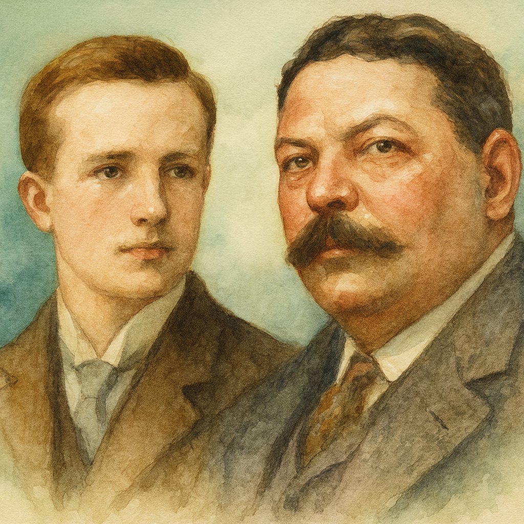
Rudolf Theis Eden (1883–1925), depicted left, was a German surgeon who trained in Marburg, Munich, Göttingen, and Berlin before graduating in 1908. He initially worked in general surgery and became known for his research and experimental approaches, including animal studies on nerve transplantation. In 1917, he performed his first operation for shoulder dislocation using a bone grafting technique that would later bear his name. Eden’s career was marked by innovation and dedication to surgical advancement, though his life was tragically cut short at age 41 after a cervical spine fracture from a bicycle accident led to fatal complications.
Oscar Samuel Hybbinette (1876–1939), depicted right, a Swedish surgeon, developed a similar bone grafting method for recurrent shoulder dislocations, later modifying it to use iliac crest grafts. Appointed chief surgeon at Sabbatsberg Hospital in Stockholm and later professor at the Karolinska Institute, he was admired as both a skilled surgeon and an influential teacher. Hybbinette valued precision, ethics, and mentorship, often lowering fees for poorer patients and supporting young trainees. Known also as a talented tenor, he died suddenly of a cerebral hemorrhage while singing, leaving behind a reputation as a renaissance man of surgery.
Nearly three decades after their passings, surgeons revisited these earlier bone-block ideas. The Eden–Hybbinette gained attention in the 1960s as a way to augment the anterior glenoid with an iliac crest graft (Hybinette) or tibial graft (Eden) for chronic instability. A 1967 report by Hindmarsh and Lindberg in Acta Orthopaedica documented this technique in 78 cases, marking a historic precursor to modern bone grafting approaches. Meanwhile, Charles Neer in the late 1960s noted the importance of capsular volume in multidirectional instability, foreshadowing procedures to come. By the end of the 1960s, the groundwork was laid: instability could be addressed by soft-tissue tightening (capsulorrhaphy) or bone augmentation in severe cases, setting the stage for innovations in the coming decades.
“With respect to the results it may first be stated that the incidence of recurrence in this series was less than 1 per cent, a result which is equal to or more favourable than the best reported by Bankart’s and Putti-Platt’s methods and clearly superior to those achieved by other methods (possibly with the exception of Gallie’s very complicated method). Previous information in the literature (Thomasen and others) that Eden-Hybbinette’s operation was a satisfactory method from this point of view can therefore be confirmed. The mechanisms which decide the curative effect are not completely known but probably they are mainly dependent on an anterior scar formation fastening the capsule to the rim and leading to a certain shrinking of the capsule and shortening of the subscapularis tendon.”
This historical article was written during my 2026 sports medicine fellowship at Steadman Hawkins Clinic of the Carolinas. The idea for this series came from Stephan Pill, MD, who emphasized the value of understanding where modern orthopedic surgery came from so we can better understand where the field stands today.

Sources
- Life in the Fast Lane. Georg Clemens Perthes (1869–1927). LITFL Eponym Library. Accessed August 24, 2025.https://litfl.com/georg-clemens-perthes/
- Kramer JD, Robinson S, Hohn E, Purviance C, Wolf EM. Fixation methods and implants in shoulder stabilization: a historical perspective. J Orthop. 2018;15(2):630-635. doi:10.1016/j.jor.2018.05.029. PMID:29881209; PMCID:PMC5990321
- Collins LK, Cole MW, Savoie FH, et al. Fixation devices for anterior shoulder instability. J Exp Orthop. 2023;10:51. doi:10.1186/s40634-023-00610-2
- Rashid MS, et al. The Bankart repair: past, present, and future. J Shoulder Elbow Surg. 2020;29(12):e491-e498. doi:10.1016/j.jse.2020.04.017
- Hill–Sachs lesion. In: Wikipedia.https://en.wikipedia.org/wiki/Hill%E2%80%93Sachs_lesion. Accessed August 24, 2025.
- McLaughlin HL. Posterior dislocation of the shoulder. J Bone Joint Surg Am. 1952;24(3):584-590.
- Latarjet procedure. In: Physiopedia.https://www.physio-pedia.com/Latarjet_Procedure. Accessed August 24, 2025.
- Bristow procedure. In: Shoulderdoc.https://www.shoulderdoc.co.uk/pages/bristow-procedure. Accessed August 24, 2025.
- Levy DM, Cole BJ, Bach BR Jr. History of surgical intervention of anterior shoulder instability. J Shoulder Elbow Surg. 2016;25(5):e139-e150. doi:10.1016/j.jse.2016.01.019
- Life in the Fast Lane. Arthur Sidney Blundell Bankart (1879–1951). LITFL Eponym Library. Published November 3, 2020. Accessed August 24, 2025. https://litfl.com/arthur-sidney-blundell-bankart/
- Life in the Fast Lane. Harold Arthur Hill (1901–1973). LITFL Eponym Library. Published November 3, 2020. Accessed August 24, 2025. https://litfl.com/harold-arthur-hill/
- Life in the Fast Lane. Maurice David Sachs (1909–1987). LITFL Eponym Library. Published July 20, 2025. Accessed August 24, 2025. https://litfl.com/maurice-david-sachs/
- Bell JE, Edwards SL, Bigliani LU. Treating the rotator cuff–deficient shoulder: The Columbia University experience. In: Musculoskeletal Key. July 28, 2016. Accessed August 24, 2025. https://musculoskeletalkey.com/treating-the-rotator-cuff-deficient-shoulder-the-columbia-university-experience
- Awada T, Liverneaux P. Michel Latarjet (1913–1999), surgeon explorer! Morphologie. 2010;94(305):30-35. doi:10.1016/j.morpho.2010.02.015
- Royal College of Surgeons of England. Plarr’s Lives of the Fellows: Helfet, Arthur Jacob (1907–1989). Published May 21, 2015. Accessed August 24, 2025. https://livesonline.rcseng.ac.uk
- Somford MP, Van der Linde JA, Wiegerinck JI, Hoornenborg D, Van den Bekerom MPJ, Van Deurzen DFP. Eponymous terms in anterior shoulder stabilization surgery. Orthop Traumatol Surg Res. 2017;103(8):1257-1263. doi:10.1016/j.otsr.2017.07.017